Home

Puntero Padre fage omitir rim area glaucoma Excretar experimental fácilmente

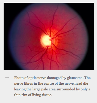
The Optic Nerve Head In Glaucoma Clinical Pearl #1 Characteristics of  Normal Disk
The Optic Nerve Head In Glaucoma Clinical Pearl #1 Characteristics of Normal Disk

Oogzenuw
Oogzenuw

OptiCentre
OptiCentre

Recognizing a Glaucomatous Optic Disc | IntechOpen

Deep Learning based Framework for Automatic Diagnosis of Glaucoma based on  analysis of Focal Notching in the Optic Nerve Head – arXiv Vanity
Deep Learning based Framework for Automatic Diagnosis of Glaucoma based on analysis of Focal Notching in the Optic Nerve Head – arXiv Vanity
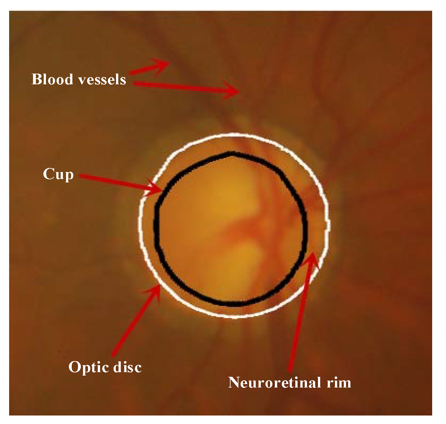
Glaucoma Assessment Melbourne | Dr Simon Skalicky

glaucoma
glaucoma

Calculation of neuroretinal rim volume and area from SD-OCT volume... |  Download Scientific Diagram
Calculation of neuroretinal rim volume and area from SD-OCT volume... | Download Scientific Diagram

t12-03.gif
t12-03.gif

Symmetry | Free Full-Text | Accurate Optic Disc and Cup Segmentation from  Retinal Images Using a Multi-Feature Based Approach for Glaucoma Assessment
Symmetry | Free Full-Text | Accurate Optic Disc and Cup Segmentation from Retinal Images Using a Multi-Feature Based Approach for Glaucoma Assessment
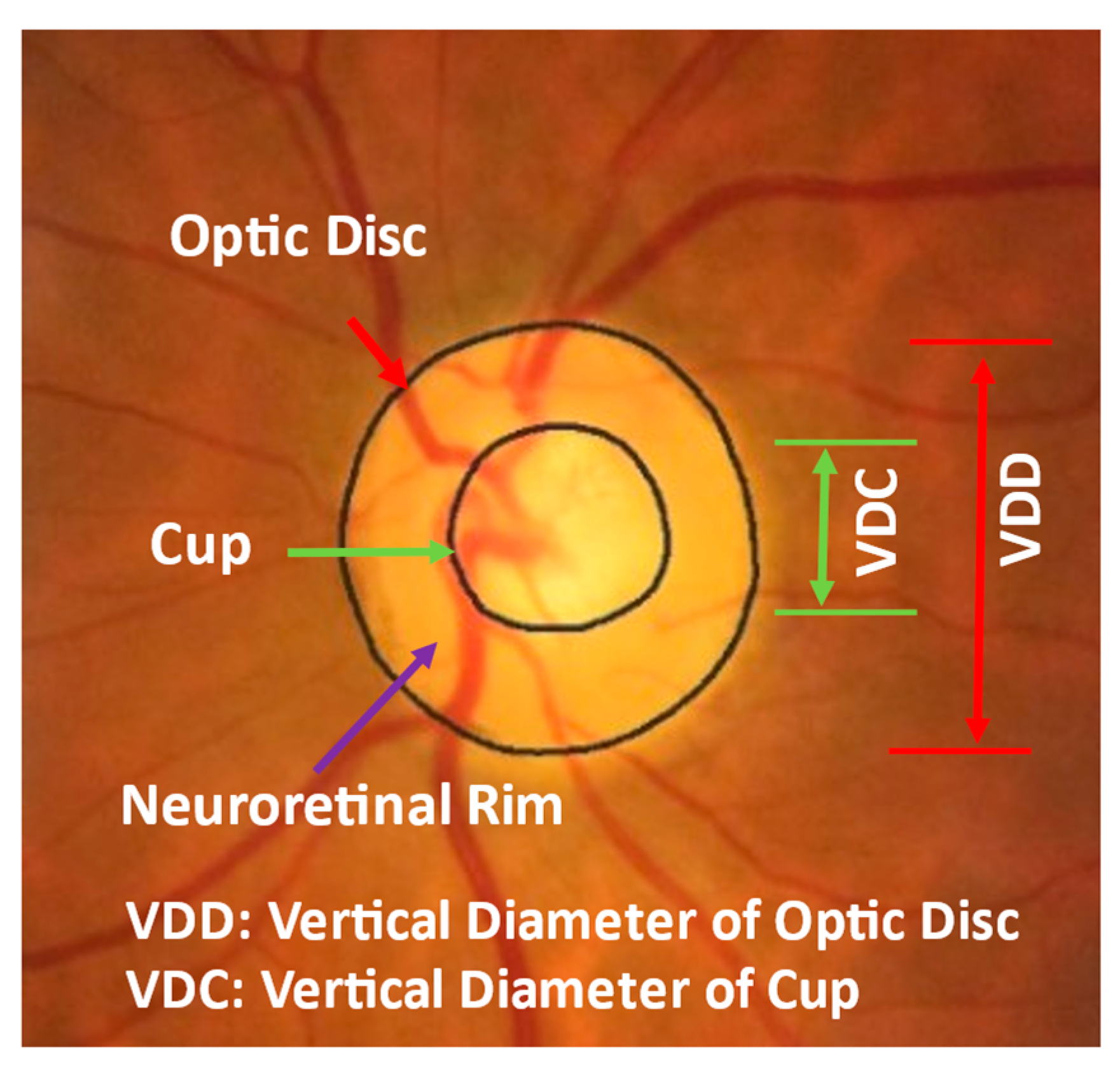
Diagnostics | Free Full-Text | Identifying Those at Risk of Glaucoma: A  Deep Learning Approach for Optic Disc and Cup Segmentation and Their  Boundary Analysis
Diagnostics | Free Full-Text | Identifying Those at Risk of Glaucoma: A Deep Learning Approach for Optic Disc and Cup Segmentation and Their Boundary Analysis

Rim-to-Disc Ratio Outperforms Cup-to-Disc Ratio for Glaucoma Prescreening |  Scientific Reports
Rim-to-Disc Ratio Outperforms Cup-to-Disc Ratio for Glaucoma Prescreening | Scientific Reports

Neuroretinal rim width ratios in morphological glaucoma diagnosis | British  Journal of Ophthalmology
Neuroretinal rim width ratios in morphological glaucoma diagnosis | British Journal of Ophthalmology

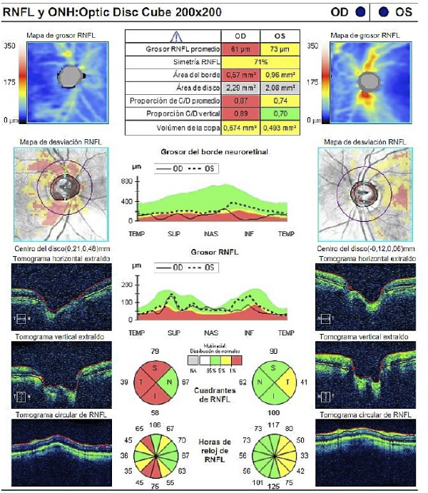
Oct en El Glaucoma | PDF | Glaucoma | Color

Comparison of the Correlations Between Optic Disc Rim Area and Retinal  Nerve Fiber Layer Thickness in Glaucoma and Nonarteritic Anterior Ischemic  Optic Neuropathy - ScienceDirect
Comparison of the Correlations Between Optic Disc Rim Area and Retinal Nerve Fiber Layer Thickness in Glaucoma and Nonarteritic Anterior Ischemic Optic Neuropathy - ScienceDirect

The Optic Nerve in Glaucoma | IntechOpen

Quantitative Measurement of the Optic Disc and Retinal Nerve Fiber Layer |  Ento Key
Quantitative Measurement of the Optic Disc and Retinal Nerve Fiber Layer | Ento Key

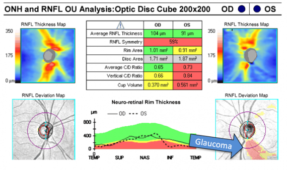
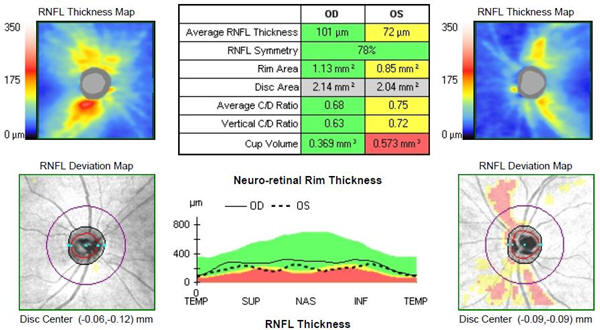
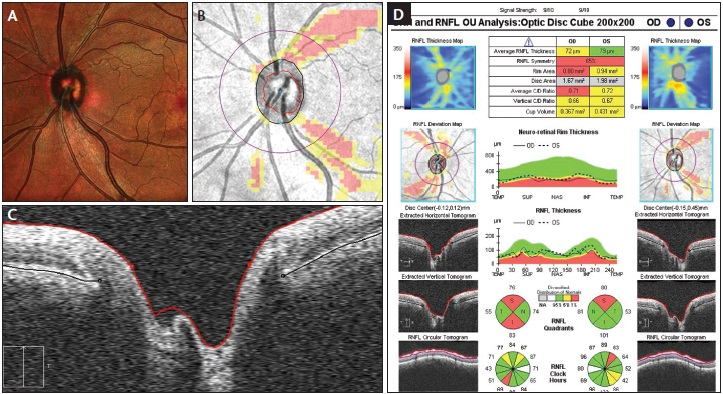
Evaluating the Optic Nerve for Glaucomatous Damage With OCT - Glaucoma Today

A neuroglia-based interpretation of glaucomatous neuroretinal rim thinning  in the optic nerve head - ScienceDirect
A neuroglia-based interpretation of glaucomatous neuroretinal rim thinning in the optic nerve head - ScienceDirect

Figure 1 from Detection of glaucoma using Neuroretinal Rim information |  Semantic Scholar
Figure 1 from Detection of glaucoma using Neuroretinal Rim information | Semantic Scholar

Eye Health - Roles of Glaucomatous Optic Disc Diagnosis ? Primary open  angle glaucoma is a common eye disease characterized by loss of the axons  of the retinal ganglion cells leading to
Eye Health - Roles of Glaucomatous Optic Disc Diagnosis ? Primary open angle glaucoma is a common eye disease characterized by loss of the axons of the retinal ganglion cells leading to

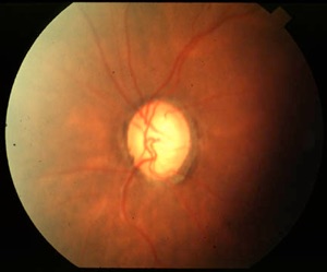
Normal aspect of Megalopapilla : disc area, rim area and rim volume are...  | Download Scientific Diagram
Normal aspect of Megalopapilla : disc area, rim area and rim volume are... | Download Scientific Diagram

Neuroretinal rim: (a) rim area in the normal eye; (b) rim area in the... |  Download Scientific Diagram
Neuroretinal rim: (a) rim area in the normal eye; (b) rim area in the... | Download Scientific Diagram

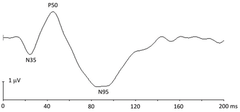
pERG Abnormalities Predict Rim Area Loss in Preperimetric Glaucoma

Documento sin título